
TL;DR
Researchers report that 10 weeks of moderate aerobic training changed the structure of nerve cells in paired clusters that help regulate the heart in rats. The changes differed between the left and right clusters, but the study does not establish whether the same pattern occurs in people or how it affects heart function.
A 10-week moderate aerobic training study in rats found that exercise changed nerve cells in paired clusters involved in regulating the heart, with different structural effects on the left and right. The University of Bristol-led team says the finding may help explain how exercise affects the nervous system, but it is not yet known whether the same changes occur in humans or how they influence heart function.

Get health and wellness essentials delivered free with Prime
- Fast, free delivery on millions of items
- Prime Video, Amazon Music and more included
- Member-only deals all year
The researchers examined the stellate ganglia, paired nerve clusters located near the lower neck and upper chest. These clusters are part of the autonomic nervous system, which controls automatic processes, including aspects of heart activity. The team included researchers from University College London, the University of São Paulo and the Federal University of São Paulo.
Using three-dimensional stereology to estimate the number, size and distribution of cells in tissue, the team found that the trained rats’ right-sided cluster contained roughly four times as many neurons as the left-sided cluster. The report describes this as a difference from the pattern in untrained rats. It also found that neurons on the left nearly doubled in size, while neurons on the right became slightly smaller.
The results, published in Autonomic Neuroscience, describe anatomical changes, not proof that exercise altered the animals’ heart rhythms or improved a heart condition. The researchers say further work is needed to establish what the structural differences mean for heart activity.
Why Left-Right Nerve Changes Matter
The findings point to a possible side-specific response in nerves that help regulate the heart. If future research establishes that similar differences occur in people and affect heart function, the results could inform studies of treatments aimed at those nerves. That possibility matters for conditions such as some arrhythmias, angina and stress-related weakening of the heart, where clinicians may seek to reduce excessive nerve signals.
Some procedures, including stellate ganglion nerve blocks and denervation, target nerve activity or pathways. The study does not show that one side should be treated over the other, nor does it test any treatment. Its potential relevance is an early research lead: understanding whether the paired clusters respond differently may eventually help researchers investigate more precisely which nerve pathways matter for particular patients.
The work also offers a biological question for exercise research. Exercise is known to place demands on the cardiovascular system, but this study suggests that adaptation may involve not only the heart muscle, but also parts of the nervous system that influence heart activity. The rat findings alone cannot establish how large or clinically meaningful that effect might be in people.
Products Worth Considering
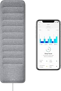
Withings Sleep Under-Mattress Sleep Tracking Pad

Withings BPM Connect Wi-Fi and Bluetooth Blood Pressure Monitor

Philips Sonicare 7300 Rechargeable Electric Toothbrush with 3 Brush Heads, Black
As an affiliate, we earn on qualifying purchases.
How the Rat Study Was Conducted
The heart is partly regulated by the autonomic nervous system, which adjusts bodily functions without conscious control. One part of this system can send signals that raise heart activity when the body needs more oxygen, including during exercise or stress. Stellate ganglia are among the nerve hubs involved in those signals, and there is one on each side.
For this study, rats completed 10 weeks of moderate aerobic training. The researchers then used stereology, a tissue-analysis method, to compare nerve-cell counts and sizes. The report does not provide a direct human comparison; its findings are based on the rats examined by the team.
Lead author Dr. Augusto Coppi, a senior lecturer in veterinary anatomy at the University of Bristol, described the paired nerve clusters as the heart’s “dimmer switch.” He said the results reveal a left-right pattern that could help explain why some treatments may work differently depending on the side targeted. That is a proposed implication, not a treatment conclusion established by this experiment.
“The findings are early-stage and in rats, so clinical studies would need to follow.”
— Dr. Augusto Coppi
Human Effects Remain Unanswered
The study was conducted in rats, not people. It remains unclear whether human stellate ganglia undergo comparable changes after regular exercise, and the report does not establish whether the observed anatomy alters heart rate, rhythm or responses to exertion.
Researchers also have not shown that the neuron-count and cell-size differences cause changes in disease risk, symptoms or treatment outcomes. The proposed links to arrhythmias, angina and stress-related heart conditions are possible future applications, not benefits demonstrated by this experiment. The study does not support changing exercise routines or medical care on the basis of its findings.
Testing Function and Human Relevance
The research team plans to investigate whether the left-right differences affect how the heart behaves at rest and during physical activity. The report also says the researchers intend to look for similar patterns in other animal models before attempting to establish whether the findings extend to humans.
For future human research, the team hopes to use noninvasive markers of nerve activity, which could offer information without surgery or tissue removal. No timeline or planned clinical study is specified in the report. Until further evidence is available, the central finding remains a structural change observed in trained rats.
Key Questions
What did the exercise study find?
After 10 weeks of moderate aerobic training, rats showed structural differences in nerve cells in the left and right stellate ganglia, clusters involved in regulating heart activity.
Was the research conducted in people?
No. The findings came from rats. Whether the same changes occur in humans has not been established.
Does the study show that exercise prevents arrhythmias?
No. The research examined nerve-cell structure and did not test whether exercise prevented or treated arrhythmias, angina or other heart conditions.
Could the findings change heart treatments?
Possibly, but that remains a future research question. The authors say understanding left-right differences could eventually inform targeted procedures, but the study does not show which side should be treated.
What research is planned next?
The team plans to study how the nerve differences relate to heart function and to look for similar patterns in other animal models and, eventually, humans. The report gives no timeline for those studies.
Source: sciencedaily.com